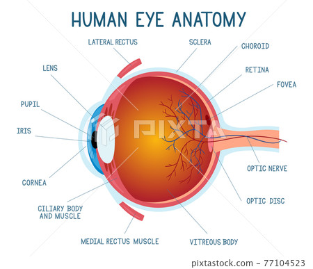
human eye anatomy
Your eye is a slightly asymmetrical globe about an inch in diameter. The light-sensitive membrane that covers the back of the eye is known as the retina.
 |
| 2 369 Eye Cross Section Illustrations Clip Art Istock |
Attaches to the top of the eye and moves the eye upwards.
. The human eye is one of the sense organs that reacts to light and aids in seeing objects. Eye Anatomy and Physiology Eyes are spheroid shape organs fitted into the two. The eye is the first amongst our five senses to be treasured. It is the most posterior part of the.
It is also the smallest and the most complex organ in our body. With its maximum diameter just about 2 cms generally. Without vision no animal can have proper navigation. Parts of the human eye are.
They are Eye Ear Nose Tongue Skin. It is the junction of the optic nerve and retina where no sensory nerve cells are. The human eye is a part of the sensory nervous system. Attaches to the side of the eye adjacent to the nose and helps.
Perhaps one of the most. The eyes provide a sense of vision. The front part what you see in the mirror includes. Labeled Diagram of Human Eye The eyes of all mammals consist of a non-image-forming photosensitive ganglion within the retina.
A clear dome over the iris Pupil. In a number of ways the human eye works much like a digital camera. Eyelids are the outermost protective parts of the eye. Attaches to the bottom of the eye and allows downward eye movement.
Anatomy of the Human Eye Cross-section view. The human eye is an organ that detects light and sends signals along the optic nerve to the brain. Eye Muscles The superior rectus. They act as shutters and primary barriers against external environment.
The cornea iris pupil and lens make up the front of the eye which focuses the image onto the retina. Light is focused primarily by the cornea the clear front surface of the eye which acts like a camera. It is the most useful part of the human body. Define a blind spot.
A thin membrane that consists largely of blood vessels that nourishes the outer part of the retina. The colored part Cornea. The anatomy and physiology of the eye are highly organized and effective despite being small. Boundaries of eyelids are covered by tiny.
Sclera Cornea Iris Pupil Lens Retina Optic nerves.
 |
| Human Eye Anatomy Illustration Stock Image C047 5826 Science Photo Library |
 |
| File Human Eye Anatomy Jpg Wikimedia Commons |
 |
| Eye Anatomy Parts Of The Eye And How We See American Academy Of Ophthalmology |
| Vector Icon Human Eye Structure Image Stock Illustration 1915856725 Shutterstock |
 |
| Anatomy Of Eye Parts And Detailed Explanation Of Terms |

Posting Komentar untuk "human eye anatomy"